Sometimes the number, shape, size or structure of teeth develops differently than expected. These differences, known as dental developmental anomalies, are usually noticed in childhood, while teeth are erupting or on a routine X-ray. In this article we explain common anomalies, how they are detected and how they are treated.
What causes dental developmental anomalies?
Teeth go through a long development process that begins before birth and continues into adolescence. Genetic factors, certain syndromes, infection or injury to a baby tooth, and some illnesses and medicines in early childhood can affect tooth development. In many cases no single cause can be identified.
Common anomalies
Anomalies of number
- Missing teeth (hypodontia): One or more permanent teeth never form. Wisdom teeth, lower premolars and upper lateral incisors are most often affected.
- Extra teeth (hyperdontia): More teeth form than normal. They are most often found between the upper front teeth and can block permanent teeth from erupting.
Anomalies of shape and size
Teeth that are smaller (microdontia) or larger than normal, peg-shaped upper lateral incisors, two teeth developing joined together (fusion) or a single tooth starting to split in two (gemination) fall into this group. These teeth are assessed for appearance, crowding and decay risk.
Structural anomalies
- Enamel defects: Enamel forms thin, rough or discoloured. Inherited forms (amelogenesis imperfecta) can affect all teeth.
- Molar-incisor hypomineralisation (MIH): Shows as white, yellow or brown patches and fragile enamel, especially on the first permanent molars and front teeth. These teeth can be sensitive and more prone to decay.
- Dentine defects: Teeth may look opaque or grey-blue and wear easily.
Eruption anomalies
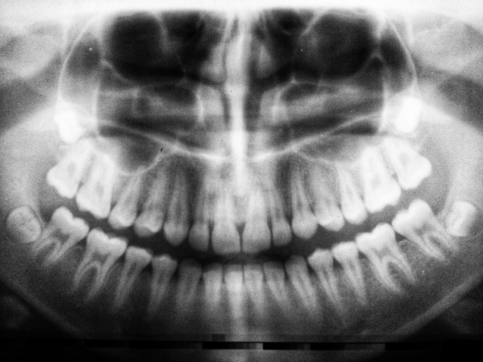
Delayed, misdirected or impacted permanent teeth are also part of monitoring tooth development. These are usually assessed with a panoramic X-ray.
How are they detected?
At the examination, the number, shape and colour of the teeth are checked. A panoramic X-ray is important because it shows whether unerupted teeth are present and where they are. Family history of similar conditions and the child’s general health history also help the assessment.
Treatment approach
Treatment is planned individually according to the type of anomaly, the child’s age and the role of the tooth. Options include measures to reduce sensitivity, fluoride and fissure sealants, reshaping with fillings, crowns for children, removing extra teeth or protecting the space of a missing tooth. In some cases a joint plan is made with orthodontics, prosthodontics or surgery.
Why early detection matters
When anomalies are noticed early, teeth can be followed in a more conservative way and any future treatment can be planned at the right time. That is why regular paediatric dental check-ups and X-ray assessment when needed are important.
Frequently asked questions
My child has white or brown spots on their teeth. Is this an anomaly?
Spots can be caused by early decay, an enamel defect or MIH, among other things. The difference is made at an examination, so a check-up is recommended when spots are noticed.
At what age are missing teeth noticed?
Missing permanent teeth are usually seen on a panoramic X-ray taken between ages 7 and 10. A baby tooth that stays for a long time can also be a sign.
Do extra teeth always need to be removed?
Not always. The decision depends on the position of the extra tooth, whether it blocks permanent teeth and the child’s age.
Are dental developmental anomalies hereditary?
Some can run in families. If there are similar conditions in your family, sharing this with your dentist helps the assessment.
Appointments for an assessment
If you have noticed something different about your child’s teeth, you can book an appointment at Korudiş’s Korupark and Ataevler branches in Bursa. See our paediatric dentistry page for more.
Korupark Branch (Osmangazi): +90 224 243 00 44 · WhatsApp +90 532 497 11 17
Ataevler Branch (Nilüfer): +90 224 503 66 00 · WhatsApp +90 552 503 66 00
Korudiş Dental Team
This article is for general information; the right treatment for you can only be decided by your dentist after an examination.